Magnetic Resonance Imaging (MRI) generation has become one of the cornerstones of cutting-edge clinical diagnostics, revolutionizing how docs visualize the human body. With its extraordinary ability to provide certain, non-invasive pix, MRI has profoundly impacted affected person care. This article will walk you via the whole thing you need to realize about MRI tech—from its foundational ideas to its modern programs and future ability.
Introduction to MRI Technology
What is MRI Technology?
MRI, or Magnetic Resonance Imaging, is a diagnostic device used to create designated pics of the frame’s inner structures using magnetic fields and radio waves. Unlike X-rays or CT scans, MRI does not depend upon ionizing radiation, making it more secure for sufferers requiring repeated imaging. It’s a favored method for inspecting soft tissues, such as the mind, muscular tissues, and organs.
The Evolution of MRI: A Brief History
The journey of MRI began inside the 1940s while researchers located nuclear magnetic resonance (NMR). By the Nineteen Seventies, scientists like Paul Lauterbur and Sir Peter Mansfield had tailored this precept for clinical imaging. Their contributions earned them a Nobel Prize in Medicine in 2003, highlighting the pivotal position of MRI in advancing healthcare.
Importance of MRI in Modern Medicine
MRI has emerged as critical in diagnosing diverse conditions, from mind tumors and spinal twine accidents to joint problems. Its potential to provide clean, three-dimensional pics lets healthcare specialists come across abnormalities early, plan remedies successfully, and reveal disease progression with precision.
How MRI Works
The Science Behind MRI
MRI is based on the interplay between magnetic fields, radiofrequency waves, and hydrogen atoms within the body. The human frame consists ordinarily of water, which contains hydrogen atoms. When uncovered to a sturdy magnetic discipline, those atoms align in a selected route. Radiofrequency pulses then disrupt this alignment, developing alerts that are captured and converted into pics.
Role of Magnetic Fields and Radio Waves
The MRI system’s magnet generates an effective magnetic field, commonly measured in teslas (T). This discipline aligns the hydrogen nuclei within the body. When the radiofrequency waves are implemented, the nuclei emit indicators as they return to their aligned kingdom. These signals are detected and processed to shape a special pix.
Understanding Resonance and Imaging
Resonance occurs when the implemented radiofrequency suits the natural frequency of hydrogen atoms. This unique interaction enhances sign exceptionality, making sure excessive-resolution images provide correct diagnostic data. The resulting pics can be tailored to emphasise one-of-a-kind tissues, which include fats, fluid, or muscle.
Key Components of an MRI Machine
An MRI machine includes 3 primary components:
The Magnet: The largest and maximum costly issue, it generates the magnetic subject necessary for imaging.
Gradient Coils: These create spatial variants in the magnetic field, permitting three-dimensional imaging.
Radiofrequency Coils: These ship and receive radio signals, taking pictures of the data wished for photo reconstruction.
Applications of MRI in Healthcare
Diagnostic Applications
MRI is primarily acknowledged for its diagnostic skills. It plays an essential role in identifying and managing conditions across numerous medical disciplines.
Neurological Imaging
MRI is well known for imaging the brain and spinal cord. It is instrumental in diagnosing conditions including strokes, multiple sclerosis, and brain tumors. Functional MRI (fMRI) is specially beneficial for mapping brain pastime and knowledge neural connections.
Musculoskeletal Imaging
Orthopedic professionals depend upon MRI to assess accidents related to ligaments, tendons, and cartilage. This makes it important for diagnosing sports activities, accidents and planning surgeries.
Therapeutic Applications of MRI
Beyond prognosis, MRI is used in healing settings. For example, it publishes interventions like biopsies and assists in making plans for radiation remedy for cancer treatment.
Advantages of MRI Technology
Non-invasive Diagnosis
MRI sticks out for being totally non-invasive, supplying a safer alternative for patients in comparison to strategies that require surgical procedure or ionizing radiation. This makes it ideal for long-time period tracking of chronic situations.
High-Resolution Imaging
The excessive degree of element furnished by means of MRI permits for specific detection of abnormalities, even in complex systems just like the brain or joints. This clarity ensures accurate diagnoses and higher patient outcomes.
Wide Range of Medical Applications
From cardiology to oncology, MRI’s versatility makes it a cornerstone of scientific imaging. Its capability to conform to unique diagnostic wishes underscores its significance in modern-day medicine.
Types of MRI Scans
Functional MRI (fMRI)
Functional MRI (fMRI) is a specialized sort of MRI that measures mind interest through detecting changes in blood drift. When a specific vicinity of the brain is extra lively, it consumes more oxygen, causing extended blood flow to that area. FMRI is commonly utilized in neuroscience research to map brain activity and recognize the functioning of different regions, specifically throughout duties like speaking, thinking, or remembering.
FMRI is also valuable in pre-surgical making plans, because it helps pick out essential regions of the mind that manipulate speech or motor features, minimizing risks for the duration of operations. Its non-invasive nature makes it an excellent tool for exploring the mysteries of human thought.
Magnetic Resonance Angiography (MRA)
MRA specializes in imaging blood vessels, making it an essential device for diagnosing vascular conditions. Unlike traditional angiography, MRA no longer requires catheter insertion or publicity to ionizing radiation. It’s usually used to stumble on aneurysms, blood clots, and arterial stenosis (narrowing of blood vessels).
This technique is particularly vital in sufferers with a history of cardiovascular sickness, as it facilitates docs to investigate the risk of strokes or coronary heart assaults and plan remedies like stenting or surgical operation.
Diffusion-Weighted Imaging (DWI)
Diffusion-weighted imaging (DWI) is a completely unique MRI approach that tracks the motion of water molecules in tissues. It is noticeably touchy to cellular modifications, making it precious for detecting early signs and symptoms of stroke or sure kinds of most cancers. For example, DWI can perceive ischemic strokes inside minutes, allowing for prompt intervention and decreasing lengthy-term damage.
This type of imaging is likewise beneficial in oncology for tracking tumor increase or comparing the effectiveness of remedies like chemotherapy or radiation.
Other Specialized MRI Techniques
MRI era has increased to include numerous specialised scans tailored to precise needs:
Cardiac MRI: Offers distinct images of the coronary heart to diagnose situations like cardiomyopathy or coronary heart valve issues.
Breast MRI: Used as a complementary device to mammography for detecting breast cancer, mainly in high-risk patients.
Spectroscopic Imaging: Analyzes chemical composition inside tissues, regularly used for brain tumors or metabolic problems.
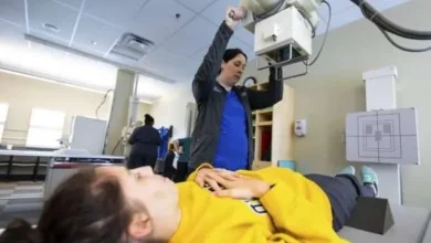
The Role of an MRI Technologist
Responsibilities and Skills of an MRI Technologist
MRI tech play a crucial role inside the imaging technique, making sure sufferers acquire correct and secure scans. Their responsibilities include:
Operating MRI machines and adjusting settings for ultimate imaging.
Preparing sufferers with the aid of explaining techniques and addressing issues.
Positioning sufferers successfully to reap clean and precise pix.
Monitoring affected person comfort and safety at some stage in the scan.
Additionally, technologists must have fantastic interpersonal abilities, technical expertise, and an intensive know-how of anatomy and imaging protocols.
Education and Certification Requirements
To become an MRI technologist, people normally complete an accomplice or bachelor’s diploma in radiologic generation, observed via specialized schooling in MRI. Certification from corporations like the American Registry of Radiologic Technologists (ARRT) is regularly required, demonstrating skill ability in MRI ideas, safety standards, and affected person care.
Continuing training is also important, as it keeps technologists updated on the modern improvements and ensures they hold their certifications.
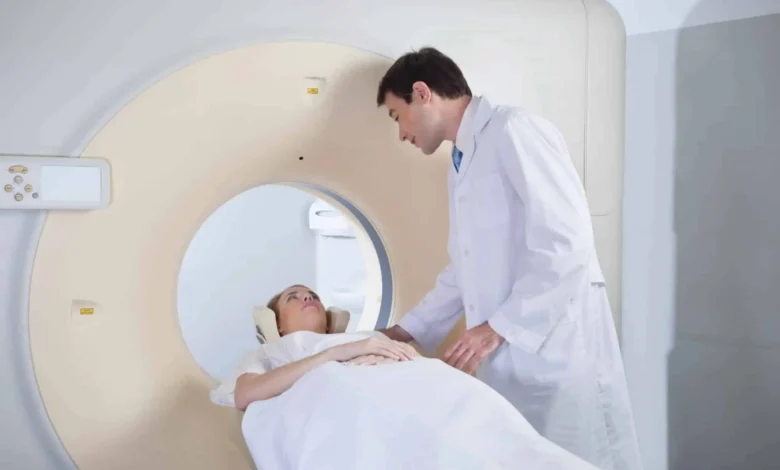
Day-to-Day Activities of an MRI Technologist
A regular day for an MRI technologist involves getting ready the imaging room, reviewing affected person facts, and appearing scans. They collaborate carefully with radiologists to make sure pix meet diagnostic standards and may assist in more complex strategies, inclusive of comparison-enhanced research.
Technologists also play a key role in calming traumatic patients, specifically those with claustrophobia, ensuring they have got superb enjoyment in the course of the scan.
MRI Safety Considerations
Risks Associated with MRI Scans
While MRI is normally safe, positive risks should be considered. The strong magnetic subject can have interaction with metal implants or devices like pacemakers, leading to headaches. Additionally, a few patients can also experience slight aspect effects from assessment sellers used at some stage in imaging, inclusive of nausea or hypersensitive reactions.
Understanding these dangers is important for ensuring affected person protection and minimizing detrimental effects.
Safety Protocols and Guidelines
MRI centers adhere to strict protection protocols to defend sufferers and groups of workers. These include:
Screening for Metal Objects: Ensuring patients do not have steel implants, earrings, or gadgets that would interfere with the magnetic area.
Monitoring Noise Levels: Providing ear protection to mitigate the loud noises produced via the device.
Contrast Agent Precautions: Using options for sufferers with allergic reactions or kidney problems.
Regular preservation of MRI machines and staff training similarly enhances protection standards.
MRI and Claustrophobia: Managing Patient Comfort
For many sufferers, the limited space of an MRI scanner can trigger claustrophobia. To address this, technologists regularly use techniques like:
Open MRI Machines: Providing an opportunity for sufferers uncomfortable with traditional scanners.
Relaxation Techniques: Encouraging deep respiration or supplying music to lessen tension.
Sedation Options: In intense instances, slight sedation can help patients whole the experiment without misery.
Recent Advancements in MRI Technology
Artificial Intelligence in MRI
Artificial intelligence (AI) is transforming MRI generation via automating image evaluation and enhancing diagnostic accuracy.
Additionally, AI-powered software reduces test instances, enhancing patient consolation and increasing the efficiency of MRI facilities.
Faster Imaging Techniques
Recent innovations have focused on dashing up MRI scans without compromising image pleasantness. Techniques like compressed sensing and parallel imaging permit for faster data acquisition, reducing scan instances extensively.
These improvements are particularly useful for pediatric or severely unwell sufferers who might also battle to stay still for lengthy periods.
Portable MRI Machines
The development of transportable MRI machines has made imaging more on hand, in particular in faraway or underserved areas. These compact devices are perfect for bedside imaging in emergency situations, presenting immediate diagnostic abilities without the need to transport sufferers.